lv enlargement radiopaedia|elevated left ventricular volume x ray : 2024-10-03 Updating. Please wait. Unable to process the form. Check for errors and try again. Perfume rating 4.07 out of 5 with 5,765 votes. Allure by Chanel is a Floral fragrance for women. Allure was launched in 1996. The nose behind this fragrance is Jacques Polge. Top notes are Lemon, Mandarin Orange, Passionfruit, Peach and Bergamot; middle notes are Honeysuckle, Jasmine, Magnolia, Freesia, Water Lily, Orange Blossom, Peony and .
0 · normal left ventricular enlargement
1 · left ventricular enlargement ultrasound
2 · left ventricular enlargement radiology
3 · left ventricular enlargement pictures
4 · left ventricular enlargement echocardiogram
5 · left ventricular enlargement ct
6 · elevated left ventricular volume x ray
7 · elevated Lv wall thickness
Malta All Inclusive Hotels. Gourmet eats, private pools, luxury spas—these value-for-money resorts have it all. Check In. — / — / — Check Out. — / — / — Guests. 1 room, 2 adults, 0 children. View map. Popular. Pool. & up. Luxury. Property types. B&Bs & Inns. Specialty lodgings. Hotels. Condos. Show more. Amenities. Free Wifi. Breakfast included.
lv enlargement radiopaedia*******The parasternal long axis and apical four-chamber views on transthoracic echocardiography are often the primary views used to gain both a qualitative and .

Updating. Please wait. Unable to process the form. Check for errors and try again.Updating. Please wait. Unable to process the form. Check for errors and try again.Annotated image demonstrating the Hoffman-Rigler sign. Left ventricular .
Dilatation of the left ventricle results to lateral and downward displacement of .On lateral view, left ventricular enlargement manifests as posterior displacement of .
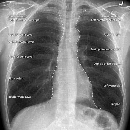
Annotated image showing the Shmoo sign for left ventricular enlargement. The .Annotated frontal and lateral chest x-ray with structures that account for the .Left ventricular hypertrophy ( LVH) is present when the left ventricular mass is .Left ventricular (LV) hypertrophy consists in an increased LV wall thickness.
Left ventricular hypertrophy (LVH) is a condition in which an increase in left ventricular mass occurs secondary to an increase in wall . LV mass assessed by echocardiography and CMR, cardiovascular outcomes, and medical practice. JACC Cardiovasc Imaging 2012;5(8):837–848.Left ventricular enlargement can be reliably identified on nongated contrast-enhanced multidetector CT when the maximum luminal diameter of the LV is greater than 5.6 cm. . Left ventricular hypertrophy was strongly associated with hard coronary heart disease events (death and myocardial infarction) over 15 years of follow-up in a . Left ventricular hypertrophy (LVH) is an unfavorable condition, which is consistently and strongly associated with significant cardiovascular morbidity and . The left ventricle is one of four heart chambers. It receives oxygenated blood from the left atrium and pumps it into the systemic circulation via the aorta. Gross anatomy. The left ventricle is conical in .Left ventricular hypertrophy (LVH): Markedly increased LV voltages: huge precordial R and S waves that overlap with the adjacent leads (SV2 + RV6 >> 35 mm). R-wave peak time > 50 ms in V5-6 with associated QRS .
The parasternal long axis and apical four-chamber views on transthoracic echocardiography are often the primary views used to gain both a qualitative and quantitative appreciation of left ventricular enlargement. Features include 4: increased left ventricular internal end-diastolic diameter (LVIDd)Left ventricular hypertrophy ( LVH) is present when the left ventricular mass is increased. It is a common condition, typically due to systemic hypertension, and it increases with age, obesity and severity of hypertension. Epidemiology. Studies have demonstrated a prevalence on echocardiography of 36-41% in hypertensive patients 1.
elevated left ventricular volume x rayLeft ventricular (LV) hypertrophy consists in an increased LV wall thickness. Left ventricular hypertrophy (LVH) is a condition in which an increase in left ventricular mass occurs secondary to an increase in wall thickness, an increase in left ventricular cavity enlargement, or both. LV mass assessed by echocardiography and CMR, cardiovascular outcomes, and medical practice. JACC Cardiovasc Imaging 2012;5(8):837–848.Left ventricular enlargement can be reliably identified on nongated contrast-enhanced multidetector CT when the maximum luminal diameter of the LV is greater than 5.6 cm. Nongated contrast-enhanced CT scan can be used to recognize LVE. Left ventricular hypertrophy was strongly associated with hard coronary heart disease events (death and myocardial infarction) over 15 years of follow-up in a contemporary ethnically diverse cohort, after adjustment for traditional risk factors and coronary artery calcium.
Left ventricular hypertrophy (LVH) is an unfavorable condition, which is consistently and strongly associated with significant cardiovascular morbidity and mortality [1]. In addition, left ventricular (LV) mass portends poor patient prognosis independent of traditional risk factors [2]. The left ventricle is one of four heart chambers. It receives oxygenated blood from the left atrium and pumps it into the systemic circulation via the aorta. Gross anatomy. The left ventricle is conical in shape with an anteroinferiorly projecting apex and is longer with thicker walls than the right ventricle.
Left ventricular hypertrophy (LVH): Markedly increased LV voltages: huge precordial R and S waves that overlap with the adjacent leads (SV2 + RV6 >> 35 mm). R-wave peak time > 50 ms in V5-6 with associated QRS broadening. LV strain pattern with ST depression and T-wave inversions in I, aVL and V5-6.lv enlargement radiopaedia The parasternal long axis and apical four-chamber views on transthoracic echocardiography are often the primary views used to gain both a qualitative and quantitative appreciation of left ventricular enlargement. Features include 4: increased left ventricular internal end-diastolic diameter (LVIDd)
Left ventricular hypertrophy ( LVH) is present when the left ventricular mass is increased. It is a common condition, typically due to systemic hypertension, and it increases with age, obesity and severity of hypertension. Epidemiology. Studies have demonstrated a prevalence on echocardiography of 36-41% in hypertensive patients 1.Left ventricular (LV) hypertrophy consists in an increased LV wall thickness.

Left ventricular hypertrophy (LVH) is a condition in which an increase in left ventricular mass occurs secondary to an increase in wall thickness, an increase in left ventricular cavity enlargement, or both. LV mass assessed by echocardiography and CMR, cardiovascular outcomes, and medical practice. JACC Cardiovasc Imaging 2012;5(8):837–848.Left ventricular enlargement can be reliably identified on nongated contrast-enhanced multidetector CT when the maximum luminal diameter of the LV is greater than 5.6 cm. Nongated contrast-enhanced CT scan can be used to recognize LVE. Left ventricular hypertrophy was strongly associated with hard coronary heart disease events (death and myocardial infarction) over 15 years of follow-up in a contemporary ethnically diverse cohort, after adjustment for traditional risk factors and coronary artery calcium. Left ventricular hypertrophy (LVH) is an unfavorable condition, which is consistently and strongly associated with significant cardiovascular morbidity and mortality [1]. In addition, left ventricular (LV) mass portends poor patient prognosis independent of traditional risk factors [2].
The left ventricle is one of four heart chambers. It receives oxygenated blood from the left atrium and pumps it into the systemic circulation via the aorta. Gross anatomy. The left ventricle is conical in shape with an anteroinferiorly projecting apex and is longer with thicker walls than the right ventricle.Left ventricular hypertrophy (LVH): Markedly increased LV voltages: huge precordial R and S waves that overlap with the adjacent leads (SV2 + RV6 >> 35 mm). R-wave peak time > 50 ms in V5-6 with associated QRS broadening. LV strain pattern with ST depression and T-wave inversions in I, aVL and V5-6. The parasternal long axis and apical four-chamber views on transthoracic echocardiography are often the primary views used to gain both a qualitative and quantitative appreciation of left ventricular enlargement. Features include 4: increased left ventricular internal end-diastolic diameter (LVIDd)
Left ventricular hypertrophy ( LVH) is present when the left ventricular mass is increased. It is a common condition, typically due to systemic hypertension, and it increases with age, obesity and severity of hypertension. Epidemiology. Studies have demonstrated a prevalence on echocardiography of 36-41% in hypertensive patients 1.Left ventricular (LV) hypertrophy consists in an increased LV wall thickness. Left ventricular hypertrophy (LVH) is a condition in which an increase in left ventricular mass occurs secondary to an increase in wall thickness, an increase in left ventricular cavity enlargement, or both. LV mass assessed by echocardiography and CMR, cardiovascular outcomes, and medical practice. JACC Cardiovasc Imaging 2012;5(8):837–848.lv enlargement radiopaedia elevated left ventricular volume x rayLeft ventricular enlargement can be reliably identified on nongated contrast-enhanced multidetector CT when the maximum luminal diameter of the LV is greater than 5.6 cm. Nongated contrast-enhanced CT scan can be used to recognize LVE.
Left ventricular hypertrophy was strongly associated with hard coronary heart disease events (death and myocardial infarction) over 15 years of follow-up in a contemporary ethnically diverse cohort, after adjustment for traditional risk factors and coronary artery calcium.
Left ventricular hypertrophy (LVH) is an unfavorable condition, which is consistently and strongly associated with significant cardiovascular morbidity and mortality [1]. In addition, left ventricular (LV) mass portends poor patient prognosis independent of traditional risk factors [2].
Allure: the very essence of elegance according to Gabrielle Chanel. Difficult to define, impossible to resist—this floral-fresh-ambery fragrance in 6 facets reveals itself in a unique way on every woman.
lv enlargement radiopaedia|elevated left ventricular volume x ray


